
- Application Article
- Download
Hooke College

- About Us
- Team
- R & D and production
- Join Us
- Contact Us
- Qualification honor


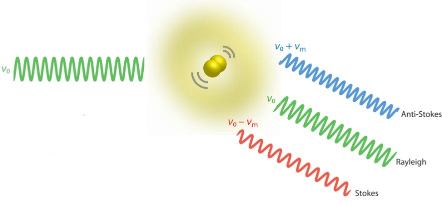
Raman spectroscopy is a lossless analytical technique, which is based on the interaction of light and material. Raman spectroscopy can provide detailed information about the chemical structure, phase and morphology, crystallinity and molecular interactions of a sample.
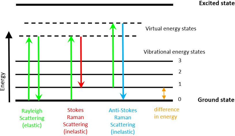
Raman is a light scattering technique. When the high-intensity incident light from a laser source is scattered by molecules, most of the scattered light has the same wavelength (color) as the incident laser, and this scattering is called Rayleigh scattering. However, a very small fraction (about 1/109) of the scattered light has a wavelength (color) different from the incident light, and the change in wavelength is determined by the chemical structure of the test sample (the so-called scattering material). This fraction of the scattered light is called Raman scattering.
Raman spectra, including peak positions and relative intensities, provide a unique chemical fingerprint that can be used to identify a substance and distinguish it from others. The actual test Raman spectrum is often very complex, and it is relatively complicated to determine the unknown object by spectral peak attribution, while searching the Raman spectrum database to find the matching result can quickly identify the unknown object.
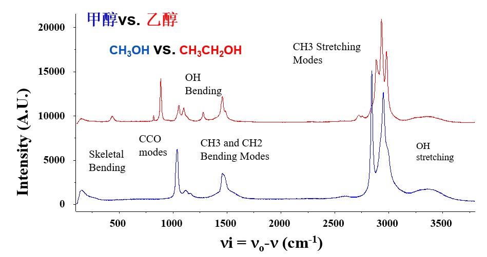
A Raman spectrum is usually composed of a number of Raman peaks, each of which represents the corresponding wavelength position and intensity of the Raman scattered light. Each spectral peak corresponds to a specific molecular bond vibration, which includes not only a single chemical bond, such as C-C, C=C, N-O, C-H, etc., but also the vibration of groups composed of several chemical bonds, such as the respiratory vibration of the benzene ring, the vibration of the long polymer chain, and the lattice vibration.
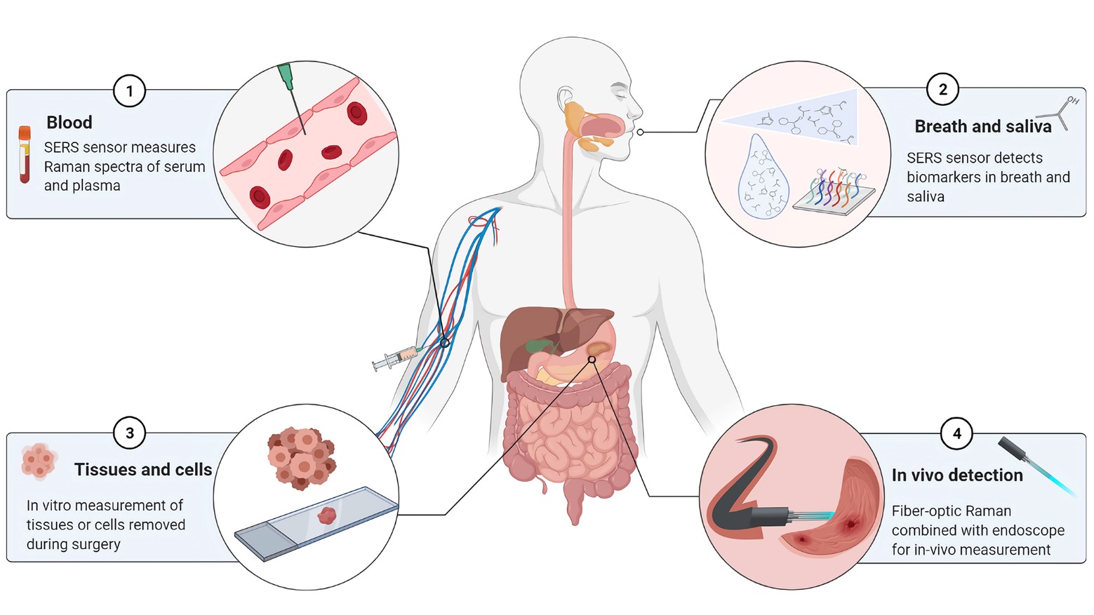
Raman spectroscopy has been widely used in biological and medical applications for more than two decades. Surface - enhanced Raman and optical tweezers Raman have achieved good results in cancer detection and organ transplant rejection detection. And the gradual maturity of instruments and software allows us to realize the transition from in vitro detection to in vivo detection. As a non-invasive test, Raman in vivo test also combines the high specificity and high sensitivity of Raman technology itself, which brings more possibilities for clinical application of Raman. Raman probe and optical fiber technology are the key to in vivo detection. This topic will mainly focus on the research of confocal Raman fiber probe, build in vivo Raman detection equipment, and mainly explore the clinical application of in vivo Raman detection in blood glucose monitoring, skin detection, surgical navigation and other aspects.
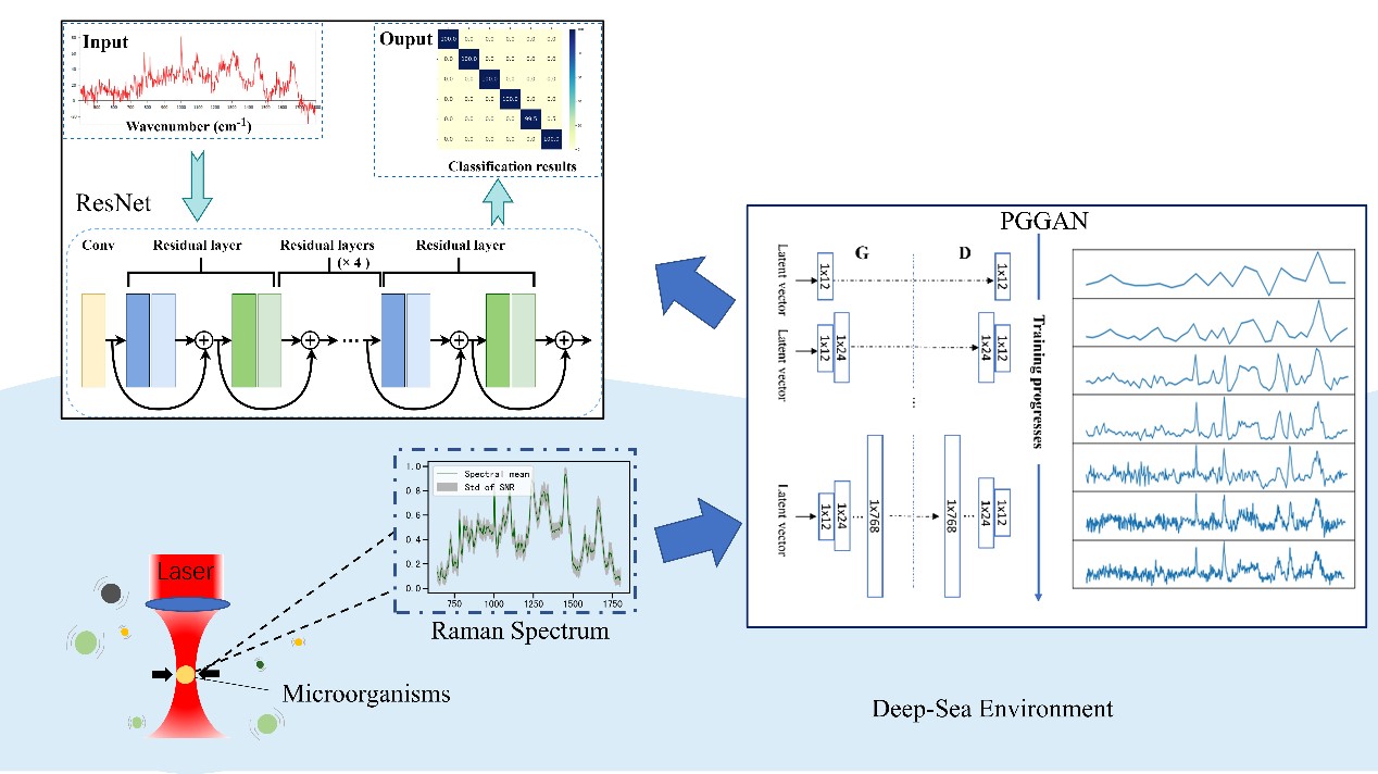
Rapid identification of Marine microorganisms is essential in Marine ecology, and Raman spectroscopy is a promising means to achieve this. Single-cell Raman spectroscopy contains biochemical characteristics of cells and can be used to identify cell phenotypes by classification models. However, traditional classification methods require a large number of reference databases, which can be very challenging when sampling in hard-to-access locations. In this case, only a few spectra are available to create classification models, which makes qualitative analysis difficult. When the spectral SNR is low, the classification accuracy will be reduced. Here, we describe a novel microbial classification method that combines optical tweezers Raman spectroscopy, progressive growth of a generative adversarial network (PGGAN), and residual network (ResNet) analysis. Using optical Raman tweezers, we obtained single-cell Raman spectra of five strains of deep-sea bacteria. We randomly selected 300 spectra from each strain as a database for training the PGGAN model. PGGAN generates a large number of high-resolution spectra similar to real data for training residual neural networks. Experimental results show that the proposed method improves the classification accuracy of machine learning and reduces the need for large amount of training data. Both methods are beneficial to analyze Raman spectra with low SNR.
Raman spectroscopy, as a kind of molecular vibration spectroscopy technology, has many advantages such as nondestructive, rapid, accurate, qualitative and quantitative detection, etc. It can obtain molecular fingerprint information, identify the properties and structures of various substances, and provide a feasible way for the detection of cancerous tissues. His current research interest is to use Raman spectroscopy technology combined with computer methods such as machine learning/deep learning to find new ways of tumor diagnosis.
In order to solve the problem that the grating diffraction efficiency is affected by the polarization state, which leads to the low overall diffraction efficiency, this paper combines the polarized light modulation technology, that is, using the polarization conversion element and special light path to design the light incident grating with a single polarization state, so as to achieve the purpose that the grating diffraction efficiency is independent of the polarization. The optical path design of polarized light modulation will further improve the flux of the spectrometer system to achieve a better spectral signal-to-noise ratio. On the other hand, the system resolution is improved by ZEMAX simulation to optimize the system aberration.
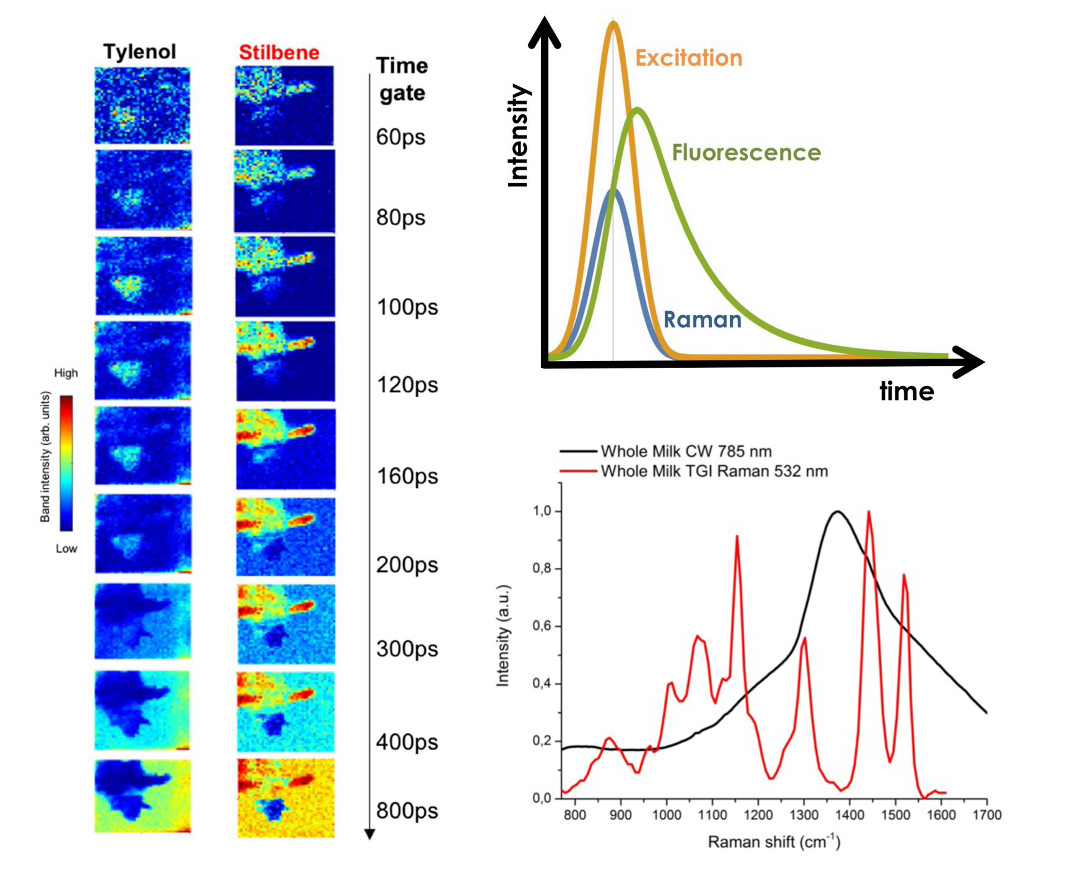
When the concept of Raman light came into being, many Raman researchers were troubled by the spectral problems caused by the fluorescence of materials. People can't completely eliminate the influence of fluorescence either by using shorter and longer wavelength laser as excitation light or SERS technology. Therefore, how to suppress the influence of fluorescence on Raman has always been a hot topic in Raman research.
Time gating Raman concept first appeared in the 1980 s, it was found that when the pulse laser action on samples, Raman light and fluorescence generation time have difference, can make use of this feature can suppress fluorescence, when laser irradiation to sample, fluorescence is not immediately can produce, it is always better than the Raman photonic delay for a period of time. When the laser stops shining, the fluorescence continues for a few nanoseconds or even hundreds of nanoseconds. Therefore, if the ultra-short pulse laser is used as the excitation light source and only the scattered light in the excitation pulse is collected, most of the fluorescence will not be received by the photodetector, so as to achieve the effect of inhibiting fluorescence. However, due to the lack of reliable photon detection equipment at that time, time-gated Raman has not been able to achieve a great breakthrough. It was not until the emergence of stable and sensitive SPAD after 2010 that time-gated Raman had the opportunity to achieve faster development.
Research the spatial heterogeneity of microbial cell, improving microbial single-celled detection method based on the Raman spectra, solve the measurement of a single microbial cell sizes and confocal Raman laser equipment spot is not easy to change this challenge, true whole-cell Raman fingerprint spectrum, coupled with perfect statistical analysis tools, Raman spectroscopy can be used to identify microorganisms more accurately at the species level.
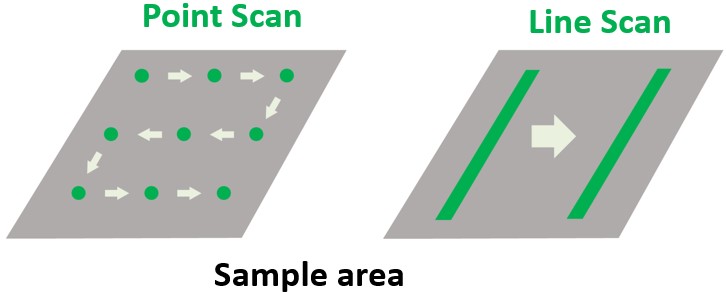
Line scanning Raman spectroscopy is a fast detection and imaging technology. Raman spectroscopy imaging technology generates a detailed chemical image based on the Raman spectrum of a sample, resulting in a pseudo-color image that reflects the composition and structure of the material. The traditional Raman spectral imaging technology usually needs to collect samples one by one, which requires relatively long acquisition time. Line scanning Raman spectroscopy uses a line as the excitation spot, whose width converges to the diffraction limit width. A single acquisition can obtain the spectral information of the line spot covering all sample points. Compared with the traditional point detection Raman spectroscopy technology, the detection speed can be increased by about two orders of magnitude.

+86-431-81077008
+86-571-86972756

Building 3, Photoelectric Information Industrial Park, No.7691 Ziyou Road, Changchun, Jilin, P.R.C
F2006, 2nd Floor,South Building, No. 368 Liuhe Road, Binjiang District, Hangzhou, Zhejiang,P.R.C

sales@hooke-instruments.com

COPYRIGHT©2022 HOOKE INSTRUMENTS LTD.ALL RIGHTS RESERVED 吉ICP备18001354号-1